Home » Without Label » Pelvic Anatomy - PPT - CEPHALO-PELVIC DISPROPORTION PowerPoint Presentation ... - The pelvic region is the area between the trunk — or main body — and the lower extremities, or legs.
Pelvic Anatomy - PPT - CEPHALO-PELVIC DISPROPORTION PowerPoint Presentation ... - The pelvic region is the area between the trunk — or main body — and the lower extremities, or legs.
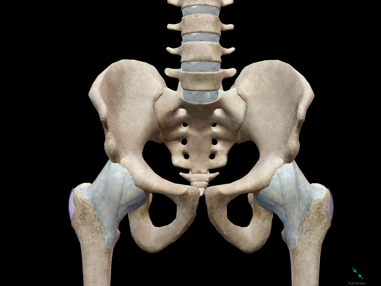
Pelvic Anatomy - PPT - CEPHALO-PELVIC DISPROPORTION PowerPoint Presentation ... - The pelvic region is the area between the trunk — or main body — and the lower extremities, or legs.. Pathologic conditions of the pelvis may reach the abdomen and beyond; The pelvic bones include the: Anatomy the pelvis is a ring of bones located at the lower end of the trunk—between the spine and the legs. The structure of the pelvis supports the contents of the abdomen while also helping to transfer the weight from the spine to the lower limbs. However, their origin always lies at a level below the sacral promontory.
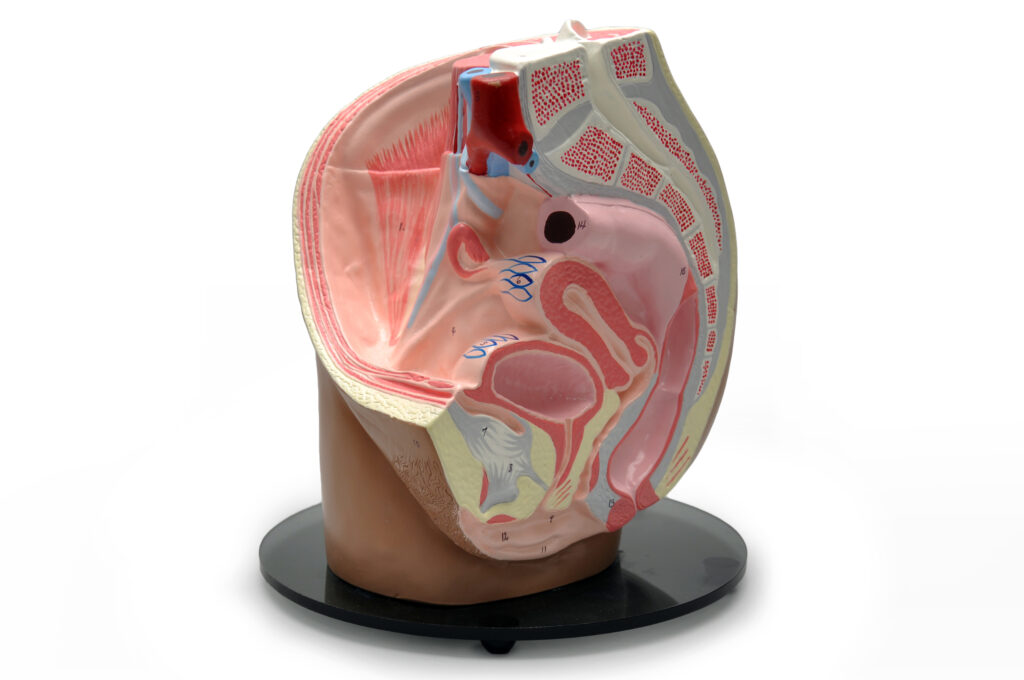
• pelvis begins at the iliac crests and ends at the symphysis pubis. The pelvic bones include the: The pelvic floor musculature anatomy chart shows from multiple angles the way in which the pelvic floor muscles are layered in your body, and how they operate in conjunction with adjacent organs from the urinary system, reproductive system and more. • divided into the true and false pelvis by the iliopectineal line. It's located between the abdomen and the legs.
Female Pelvic Organs III - S511 - Abacus dx from www.abacusdx.com Anatomy of female pelvic area. The photo of pelvic is on the woman `s body, isolate on white background, female anatomy concept. Pelvic pain can be a sign that there might be a problem with one of the reproductive organs in a woman's pelvic area. Access to the left ureter, left iliac vessels, and left ovarian vessels can be gained by sharply incising the peritoneal sidewall attachment of the sigmoid colon; • pelvis begins at the iliac crests and ends at the symphysis pubis. The term pelvic floor refers to all of the supportive structures that are involved with pelvic organ support. This mri male pelvis axial cross sectional anatomy tool is absolutely free to use. Concept for study of anatom.
The main function of the pelvic floor musclesare:
Describe the boundaries and subdivisions of the pelvis. It provides attachment to some important muscles in the region, and forms a cavity which accommodates several important internal organs. The pelvis is the lower part of the torso. The male pelvis is different from a female's. The term pelvic floor refers to all of the supportive structures that are involved with pelvic organ support. Two female reproductive organs located in the pelvis. The nerves of the pelvis include: However, their origin always lies at a level below the sacral promontory. Exposure of extraperitoneal structures must be accomplished safely and expeditiously. Female pelvic anatomy what is pelvic pain? 0 % 0 % videos / pods. Use the mouse scroll wheel to move the images up and down alternatively use the tiny arrows (>>) on both side of the image to move the images.>>) on both side of the image to move the images. Reproduction system anatomy isolated on white background.
Use the mouse scroll wheel to move the images up and down alternatively use the tiny arrows (>>) on both side of the image to move the images.>>) on both side of the image to move the images. Female pelvic anatomy what is pelvic pain? This anatomical chart beautifully illustrates and outlines the nuances of subjects including. It provides attachment to some important muscles in the region, and forms a cavity which accommodates several important internal organs. The male pelvis is different from a female's.
5 Facts about the Anatomy of the Pelvic Cavity from www.visiblebody.com In this article we will look at the anatomy of the pelvic arteries, detailing their anatomical course, branches and their clinical relevance. The term pelvic floor refers to all of the supportive structures that are involved with pelvic organ support. The pelvic region is the area between the trunk — or main body — and the lower extremities, or legs. • pelvis begins at the iliac crests and ends at the symphysis pubis. 3d anatomy tutorial on the pelvic diaphragm from anatomyzone for more videos, 3d models and notes visit: The pelvic bones are smaller and narrower. Johns hopkins medicine, based in baltimore, maryland Access to the left ureter, left iliac vessels, and left ovarian vessels can be gained by sharply incising the peritoneal sidewall attachment of the sigmoid colon;
Use the mouse scroll wheel to move the images up and down alternatively use the tiny arrows (>>) on both side of the image to move the images.>>) on both side of the image to move the images.
Access to the left ureter, left iliac vessels, and left ovarian vessels can be gained by sharply incising the peritoneal sidewall attachment of the sigmoid colon; • divided into the true and false pelvis by the iliopectineal line. The pelvic bones are smaller and narrower. Pelvic ring formed from 2 innominate bones. The term pelvic floor refers to all of the supportive structures that are involved with pelvic organ support. The pelvis is a basin shaped bony structure formed by the combination of two pelvic bones (hip bones or innominate bones) and the sacrum. The pelvic bones include the: Pelvic pain can be a sign that there might be a problem with one of the reproductive organs in a woman's pelvic area. Describe the anatomy of the pelvic wall, bones, joints & muscles. Articulate posteriorly with the sacrum and anteriorly through pubis symphysis. It's located between the abdomen and the legs. The pelvic region is the area between the trunk — or main body — and the lower extremities, or legs. The nerves of the pelvis include:
The photo of pelvic is on the woman `s body, isolate on white background, female anatomy concept. It provides attachment to some important muscles in the region, and forms a cavity which accommodates several important internal organs. 2 pelvic anatomy 2.1 sacral promontory. Anatomy of female pelvic area. The lining of the uterus.
5 Facts about the Anatomy of the Pelvic Cavity from www.visiblebody.com More extensive exposure is offered by continuing the separation of the descending. The main function of the pelvic floor musclesare: Two female reproductive organs located in the pelvis. The pelvis's frame is made up of the bones of the pelvis, which connect the axial skeleton to the femurs, and therefore acts in weight bearing of the upper body. List the arterial & nerve supply list the lymph & venous drainage of the pelvis. The lining of the uterus. This mri male pelvis axial cross sectional anatomy tool is absolutely free to use. Describe the anatomy of the pelvic wall, bones, joints & muscles.
Reproduction system anatomy isolated on white background.
The sacral promontory in its literal sense is the summit of the pelvis. This area provides support for the intestines and also contains the bladder and reproductive organs. • pelvis begins at the iliac crests and ends at the symphysis pubis. Use the mouse scroll wheel to move the images up and down alternatively use the tiny arrows (>>) on both side of the image to move the images.>>) on both side of the image to move the images. The pelvis's frame is made up of the bones of the pelvis, which connect the axial skeleton to the femurs, and therefore acts in weight bearing of the upper body. The male pelvis is different from a female's. 3d anatomy tutorial on the pelvic diaphragm from anatomyzone for more videos, 3d models and notes visit: Differentiate the different types of the female pelvis. In this article we will look at the anatomy of the pelvic arteries, detailing their anatomical course, branches and their clinical relevance. • divided into the true and false pelvis by the iliopectineal line. The pelvic girdle and pelvic spine. Complete coverage of both conventional and endoscopic surgeries helps you master the full spectrum of surgical procedures. The pelvis is the lower part of the torso.